Application
X-ray technology
Application
X-ray technology
Razor-sharp images thanks to myonic X-ray tube bearings
X-rays not only help to identify a possible bone fracture or an infected tooth root. Even the smallest blood clots or tumors in the brain can be detected with a CT scan.
The top quality of myonic bearings ensures precise and low-vibration running and therefore a razor-sharp X-ray image. But X-rays are not only used for diagnosis, they are also successfully used in the fight against tumors.

A P P L I C A T I O N E X A M P L E
Precisely irradiated
CyberKnife system – precise and innovative radiotherapy treatment
The CyberKnife system is the first and only robotic whole-body radiosurgery system that offers non-operable treatment options for a variety of tumors including prostate, lung, spine, liver, pancreas and other extracranial tumors.
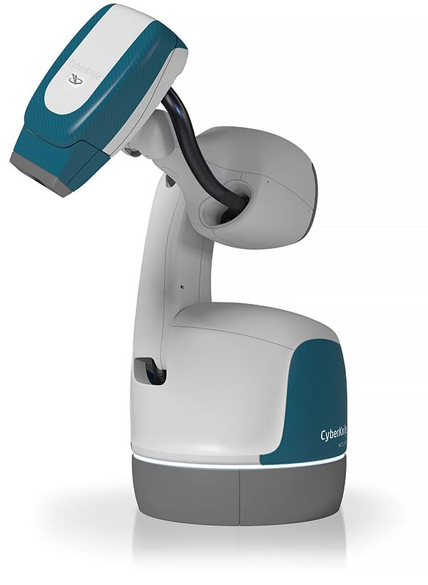
The head of the CyberKnife system contains the InCise(tm) multileaf collimator system, which is used to adapt the required radiation to the shape of the tumor, thereby minimizing the exposure of the surrounding healthy areas to radiation.
The InCise(tm) multileaf collimator system from our customer Accuray (USA) consists of a large number of lead plates (leaves) that can be constantly repositioned in real time during treatment.
This means that the radiation area can be constantly readjusted, allowing the robot to treat the often irregularly shaped tumors from different radiation angles. It also enables doctors to efficiently and precisely irradiate tumors that move with the patient.
Available X-ray devices with rotating anodes – a brief overview
 Performance to the point

myonic bearing units for X-ray tubes are used in OEM and replacement X-ray tubes by the world’s leading manufacturers to ensure the optimum production of sharp, detailed images.
In diagnostics, e.g. when X-raying a bone fracture, high power and very short irradiation times are used to avoid movements of the patient that reduce quality. The rotating anode technology used leads to a better distribution of thermal heat, making it possible to build X-ray tubes with high performance and a long service life.
The second important characteristic of a good tube is the minimum size or sharpness of the so-called focal spot. The ball bearing system developed by myonic ensures highly accurate, low-loss and vibration-free running in every position despite temperatures of up to 500°C.
Shh, no stress
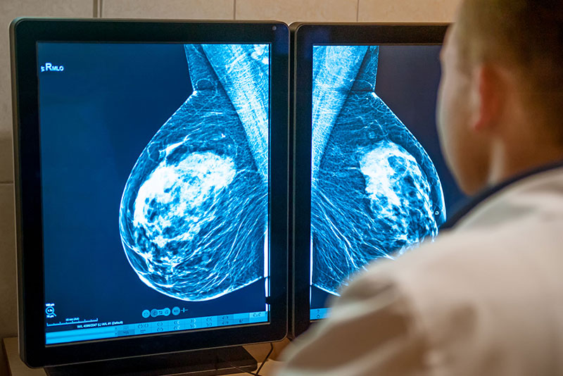
Mammography is used for the early detection of breast cancer in women without signs or symptoms of breast disease or for the diagnosis of breast disease in women with symptoms such as lumps or pain.
In this highly sensitive application, special requirements are placed on the quiet and low-vibration operation of the X-ray tube in order to ensure uniform radiation emission for optimum contrast with a low radiation dose and at the same time not to put the patient under additional stress through unnecessary noise.
Installed in the tubes of leading manufacturers worldwide, myonic bearing units have made it possible to produce many hundreds of thousands of mammograms, thus successfully contributing to the fight against breast cancer.
Going round in circles at high speed
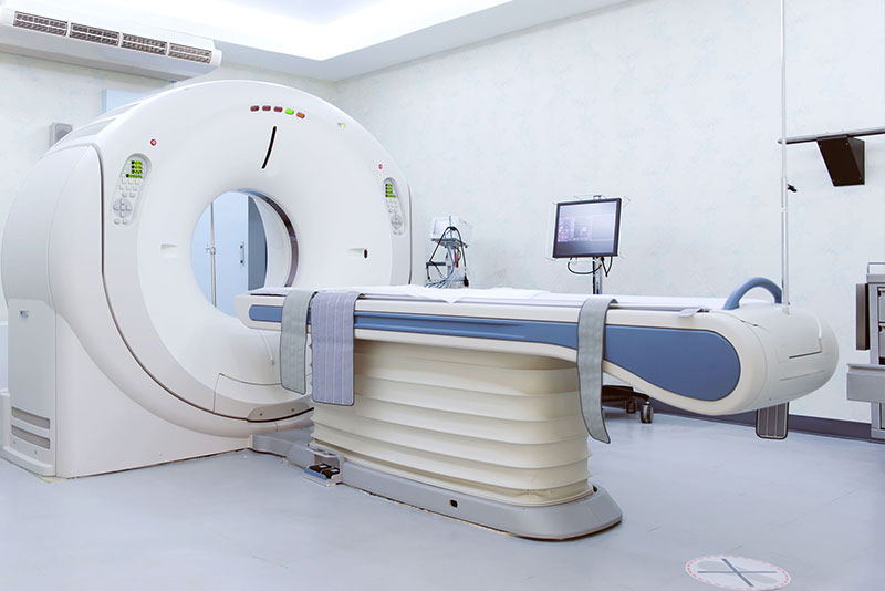
Computer tomography (CT) is a special form of X-ray. CT scans provide much more detailed images than conventional X-rays. With CT images, doctors can use special hardware and software to create very precise 3D views and any desired sectional planes of many parts of the body. Computer tomography scans are used, for example, to diagnose cerebral infarctions or bleedings, to localize lung tumors, tumors or inflammation of the abdomen, and they are also used for bone fractures or vascular problems.
To generate the required number of images, the X-ray tube unit rotates around the patient at a frequency of up to 4 times per second. The components experience an acceleration of up to 50 times the acceleration due to gravity.
The ball bearing units customized by myonic for this application can safely absorb these forces without losing their high-precision rotation or their low running noise.
Our focus is on our customers!
Numerous renowned manufacturers of X-ray equipment around the world rely on the quality of myonic products. Working closely with our customers, we are able to implement even highly complex applications.
Thanks to our close relationship with our customers, we understand the specific requirements of X-ray technology and consistently apply this experience to develop ever more powerful products.


Origin of X-rays and construction of an X-ray tube with rotating anode
The diagram shows the structure of an X-ray tube with a rotating anode. A heated filament cathode (minus) is used to generate free electrons, which are accelerated towards the anode (plus) in a vacuum by the high voltage applied – in the range of 1 kV to 10 kV.
When the electrons collide with the anode surface, two processes occur which ensure that the kinetic energy of the electrons is converted into radiation energy:
A (negatively charged) electron flies between the atoms of the anode and is deflected in the field of the (positively charged) atomic nuclei, which reduces the kinetic energy of the electron – it is decelerated (1) – and the difference in kinetic energy is converted into radiation energy. This results in so-called deceleration radiation.
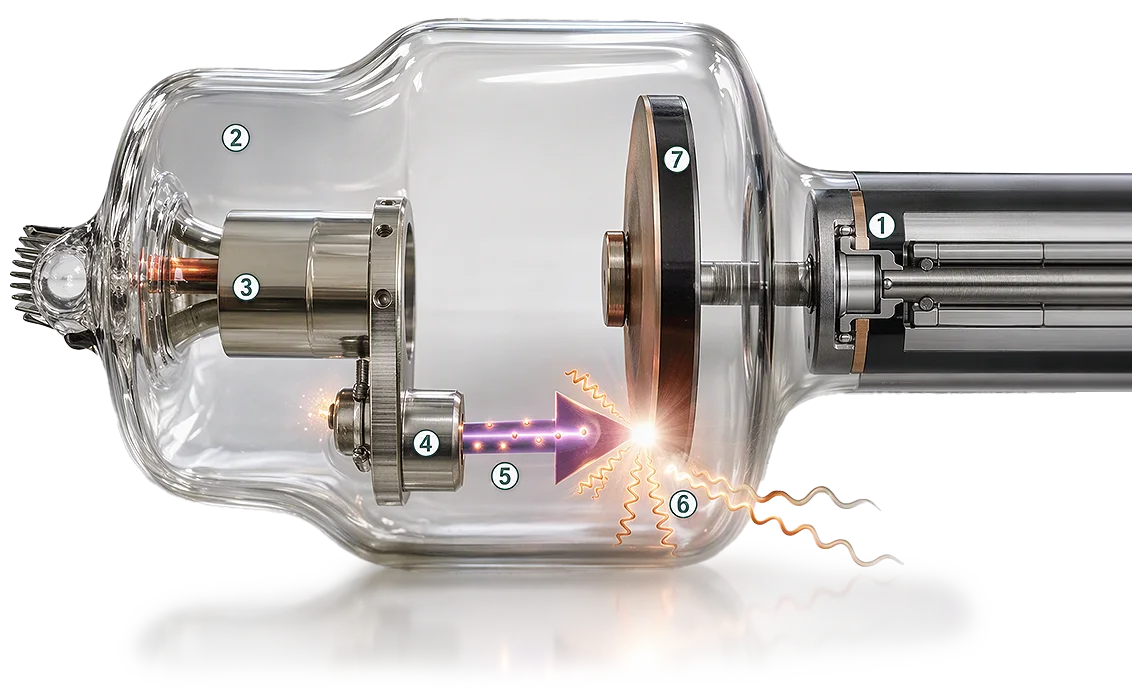
1 myonic bearing assembly 2 Vacuum 3 Cathode 4 Filament
5 Electrons 6 X-rays 7 Anode plate


Products X-ray technology
Products & Solutions
Applications